 |
Heartbeat |
||||||||||||||||||||||||||||||||||||||||||||||||||
|
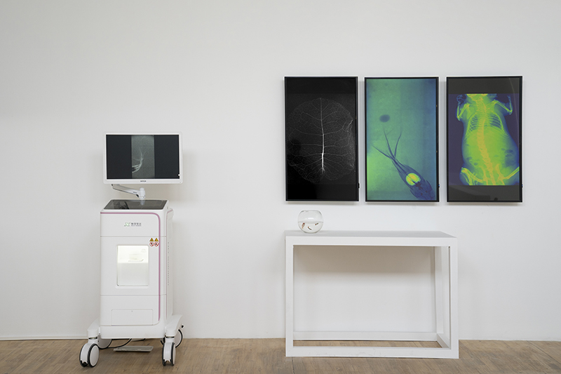
Background: In 1895, the fortuitous discovery of X-rays by German physicist Wilhelm Conrad Roentgen while studying cathode rays sparked immediate interest within the medical community. By 1896, X-rays had been employed for medical diagnostics, primarily in the detection of fractures, foreign objects, and dental examinations. The early 20th century witnessed rapid advancements in X-ray technology, with continuous improvements in X-ray tubes and equipment, leading to enhanced image quality and safety. X-rays boast widespread application in medical diagnosis, including chest radiography, fracture detection, and dental X-rays. They can provide clear visualization of skeletal structures and certain soft tissues. Additionally, X-rays are utilized as guidance during minimally invasive surgeries, such as cardiac catheterization and angioplasty. High-dose X-rays are employed in cancer therapy, aiming to destroy tumor cells by damaging their DNA. For industrial application, X-rays are employed for the detection of internal flaws within metals and welded components, including cracks, voids, and inhomogeneities. X-ray fluorescence spectroscopy (XRF) is utilized for the analysis of material composition. Airports and other venues demanding a high level of security deploy X-ray scanning machines to inspect the contents of luggage and packages. For scientific research, X-ray crystallography is employed for the determination of crystal structures, encompassing complex biological molecules such as proteins and DNA. X-ray astronomy delves into the study of high-energy celestial bodies within the cosmos, including enigmatic entities such as black holes, neutron stars, and supernova remnants. Research: X-rays are a type of ionizing radiation that can cause damage to the DNA within cells, leading to cell death or mutations. Long-term exposure to X-rays may increase the risk of developing cancer. When exposed to extremely high doses of X-ray radiation, individuals may experience acute radiation syndrome, which can manifest as skin burns, hair loss, nausea, vomiting, and organ damage. In medical imaging, healthcare professionals strive to utilize the lowest effective dose of X-rays to minimize radiation exposure to patients. A series of medical X-ray instruments developed by Shenzhen Xpect Vison Company incorporate advanced optical technology and algorithms. The photon-counting X-ray detectors and overall system exhibit numerous advantages, including zero dark noise, high spatial resolution, wide dynamic range, high linearity, and the ability to achieve energy resolution. These advancements have led to the miniaturization and real-time capabilities of X-ray machines. In the case of our real-time X-ray machine, small animals can be observed for several hours while staying alive after being irradiated with extremely low doses. The observations are presented in beautiful real-time color images. In addition to monitoring the heartbeat of animals such as hippocampi, frogs, mice, and goldfish, the most crucial aspect is the ability to observe the diffusion process of drugs after administration.
|